Why does the femoral head loose its blood supply?
1. What is Avascular Necrosis or Osteonecrosis?
2. What are the symptoms of Avascular Necrosis?
3. What are the causes of Avascular Necrosis?
4. What are the risk factors for developing Avascular Necrosis?
5. What are the investigations needed for diagnosis?
6. What are the stages of Avascular Necrosis?
7. What are the treatment options for Avascular Necrosis?
8. What complications can occur in untreated Avascular Necrosis?
9. What is Core Decompression?
10. What is Core Decompression with BMAC insertion?
11. What are the different types of hip replacements?
12. What are the different bearing options in Total Hip Replacement?
What is Avascular Necrosis or Osteonecrosis?
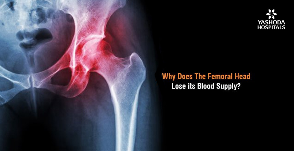
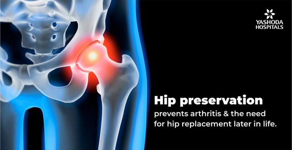
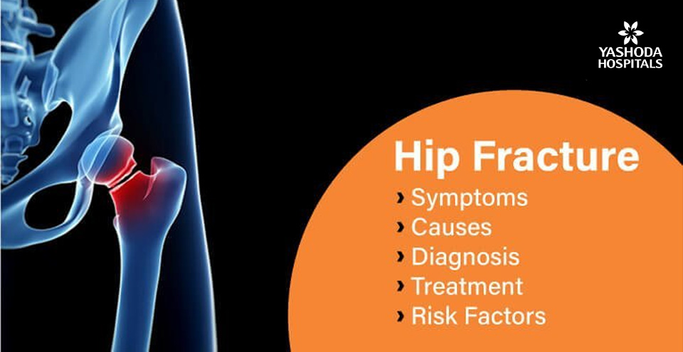
A lack of blood supply to the tissue within the bone leads to its death called Avascular necrosis or Osteonecrosis. It can sometimes lead to tiny breaks within the structure of the bone and eventually its collapse. The hip is the most commonly affected area with osteonecrosis. Other than the hip, commonly affected areas are the knee, shoulder, hand and foot.
What are the symptoms of Avascular Necrosis?
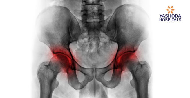
The early stages of avascular necrosis may not be associated with any symptoms. However, the affected joint may begin to hurt as the condition progresses and the person puts on weight in the affected area. The primary symptom of avascular necrosis is pain which develops gradually and can be mild or severe. Sometimes the pain may persist even on lying down. The pain is located at the centre of the groin or radiates to the area of the thigh or buttock. In certain persons, (4 to 5 out of 10) avascular necrosis may be bilateral, i.e., it develops on both hip joints.

What are the causes of Avascular Necrosis?
Osteonecrosis usually affects people between the ages of 25 and 50. The primary cause of avascular necrosis is the interruption of blood supply within the bone which can be caused by:
- Trauma or injury to a joint or bone: A traumatic injury like an accident or a condition like a dislocated joint can obliterate the adjacent blood vessels.
- Obstruction in blood vessels due to fatty deposits: The small-sized blood vessels within the bone can get blocked due to the fat deposits (lipids) which can obstruct the blood supply to the bones.
- Certain medical conditions: Blood flow to the bone can also become diminished in certain conditions like Gaucher’s disease and sickle cell anaemia. In certain cases, radiation therapy for the treatment of cancers may also lead to damage to the blood vessels and weakening of the bones.
- Unknown causes: In about one-fourth of cases diagnosed with avascular necrosis, the cause of interrupted blood flow may remain uncertain.
What are the risk factors for developing Avascular Necrosis?
Many factors can increase the likelihood of avascular necrosis such as:
- Injuries or Trauma that reduce blood flow to bones due to obliteration of feeding blood vessels.
- Use of medications like steroid use: High-dose corticosteroids used for the management of certain medical conditions like SLE, rheumatological conditions, ALL, multiple sclerosis etc. can lead to avascular necrosis probably by increasing the levels of lipid within the blood, thereby reducing blood flow.
- Alcohol excess: Excessive consumption of alcoholic drinks for a prolonged duration of time can also cause fatty deposits to form in the blood vessels, thereby increasing the tendency for obliteration.
- Use of medications like bisphosphonates: Prolonged use of medications like bisphosphonates that help in increasing the density of bones in osteoporosis, multiple myeloma and metastatic breast cancer etc may also contribute to developing osteonecrosis of the jaw as a rare complication.
- Certain medical procedures: Cancer therapy with radiations and organ transplantation like kidney transplant are also known to be associated with avascular necrosis.
Some of the medical conditions that increase an individual’s susceptibility to avascular necrosis include:
- Diabetes
- HIV/AIDS
- Pancreatitis
- Sickle cell anaemia
- Systemic lupus erythematosus
- Gaucher’s disease
What are the investigations needed for diagnosis?
The diagnosis of avascular necrosis is performed by an Orthopaedic Surgeon based on medical history, physical examination and tests. Tenderness and movements may be assessed by pressing around the joints and moving them during physical examination.
Imaging tests: Advised to determine the source of pain
- X-rays: Determine the bone changes in the later stages of avascular necrosis.
- MRI: Assess the presence of early changes in bone indicative of avascular necrosis.
What are the stages of Avascular Necrosis?
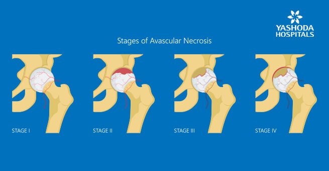
There are usually four stages of avascular necrosis.
Stage I: Progress from a normal, healthy hip to pain observed in the groin area
Stage II: Pain and stiffness
Stage III: Pain radiates to the surrounding areas like the knee
Stage IV: Pain and limp on the affected side
What are the treatment options for Avascular Necrosis?
The goal of treatment in avascular necrosis is to manage the symptoms and prevent further bone loss. Sometimes, the condition may be self-limiting and can be managed conservatively. The treatment is a combination of conservative and surgical management when required.
- Medications like pain killers and Osteoporosis medication
- Weight bearing off the joint with crutches for avascular necrosis of femoral head
- Physiotherapy and hip exercises
- Lifestyle modifications to prevent excessive alcohol consumption or treatment of alcohol dependence
Surgical options: Since the symptoms of osteonecrosis develop mostly when the condition has progressed far, the orthopaedic surgeon might recommend surgery. The commonly done surgical procedures include:
- Core decompression
- Core decompression and Bone Marrow Aspirate Concentrate (BMAC) insertion
- Total hip replacement
What complications can occur in untreated Avascular Necrosis?
Avascular necrosis is progressive in nature. If left untreated, it deteriorates with time and eventually, the bone may collapse. Progressive avascular necrosis also leads to loss of the smooth surfaces of the thereby causing severe arthritis.
What is Core Decompression?
In this procedure, the affected part of the femoral head is drilled to relieve pain and pressure in the bone. This could provide pain relief and prevent rapid progression to arthritis in select patients when there is no subchondral fracture/ collapse of the head.
What is Core Decompression with BMAC insertion?
In this procedure, the affected bone is drilled and bone marrow aspiration from Iliac crest (pelvic bone) is taken and centrifuged in a machine. This concentrate is injected into the drilled hole in the bone to help in new bone formation to strengthen the affected bone.
What are the different types of hip replacements?
The different types of hip replacements available include:
- Cemented Total Hip Replacement: The prosthesis is fixed in the bone with bone cement that acts like grout.
- Uncemented Total Hip Replacement: The prosthesis has a porous coating or hydroxyapatite coating that helps in achieving fixation in the bone.
- Hybrid Total Hip Replacement: It is the combination of the uncemented cup and cemented stem.
- Reverse Hybrid Total Hip replacement: It is the combination of the cemented cup and uncemented stem.
What are the different bearing options in Total Hip Replacement?
The different bearing options in Total Hip Replacement include:
- Ceramic – on – Ceramic
- Ceramic – on – Polyethylene
- Metal – on – Polyethylene
Which bearing surface lasts longer?
The Metal-on-Polyethylene has been the gold standard for a long duration. However, long term (>20 years) results of Ceramic bearings appear to be promising. Ceramic-on-Ceramic and Ceramic-on-Polyethylene has demonstrated to be functioning well at 20 years of follow-up studies.
References:
- Avascular necrosis, MayoClinic, https://www.mayoclinic.org/diseases-conditions/avascular-necrosis/symptoms-causes/syc-20369859. Accessed on 10th November 2020.
- Femoral Head Avascular Necrosis, Medscape, https://emedicine.medscape.com/article/86568-overview. Accessed on 10th November 2020
- Avascular Necrosis (AVN or Osteonecrosis), WebMD, https://www.webmd.com/arthritis/avascular-necrosis-osteonecrosis-symptoms-treatments. Accessed on 11th November 2020
- Femoral Head Avascular Necrosis, NCBI, https://www.ncbi.nlm.nih.gov/books/NBK546658/#. Accessed on 11th November 2020
About Author –
Dr. Praveen Mereddy, Consultant joint Replacement & Trauma Surgeon, Yashoda Hospitals – Hyderabad
MS (Ortho), DNB (Ortho), MRCS (Ed), M.Ch (Ortho), FRCS (Ortho)
Complex Primary Hip and Knee Replacement Surgery, Partial Knee Replacement Surgery, Treatment of Painful/Unstable/Failed (Loosening/infection) Primary Joint Replacements, Complex, complicated fractures and Pelvi-acetabular trauma









 Appointment
 Second Opinion
 WhatsApp
 Call
Call More
More


