Lumbar and Cervical Spinal Stenosis
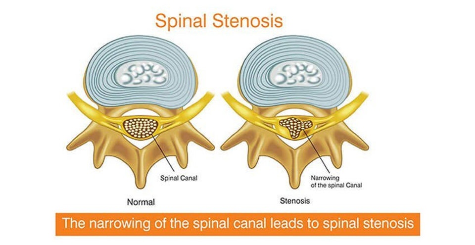
The narrowing of the spinal canal leads to spinal stenosis
In some people there is narrowing of open spaces within the spine. This condition which is described as spinal stenosis leads to pressure on the spinal cord and nerves of arms and legs. Spinal stenosis occurs in the lower back and neck regions. It is marked by pain, tingling, numbness, muscle weakness, prevalence of disturbed bladder and bowel functions. Spinal stenosis is primarily caused by osteoporosis. Treatment for this condition includes drugs, physical therapy and surgery.
CAUSES
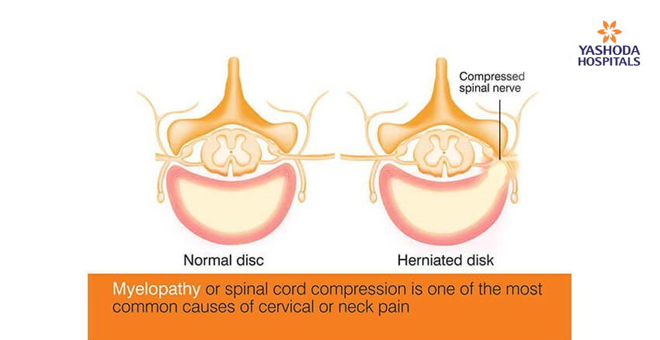
Spinal stenosis may occur due to many factors viz. overgrowth of bone, herniated discs, and thickened ligaments. Tumors and spinal injuries may also lead to spinal stenosis. In this condition, there is abnormal growth of the bones and tumors within the spinal cord reduce the space between the vertebrae and spinal cord.
SYMPTOMS
Symptoms are dependent on the location of spinal stenosis. If the neck region is affected the symptoms are evident as numbness and tingling of leg, foot, arm or hand. Walking and balance are affected. There is tingling in the hands. In its extreme, incontinence is seen with poor bladder and bowel function. When the lower back is affected, symptoms are evident as pain and cramps in the legs.
RISK FACTORS AND COMPLICATIONS
Those who are 50 years and above are greatly prone to spinal stenosis. However, youngsters may also show symptoms of spinal stenosis, which is majorly due to genetic factors relating to bone and muscle development. The complications of spinal stenosis are evident as numbness of the hands and legs, intense fatigue, balance problems, chronic incontinence and paralysis.
TESTS AND DIAGNOSIS
The doctor may enquire about the age, life style and health history of the patient. All the symptoms experienced by the person are noted. To confirm on spinal diagnosis, the doctor may advise for imaging tests likewise X-rays, Magnetic resonance imaging (MRI), and CT Myelogram. X-ray images helps to know about the bone changes, and narrowing of the space within the spinal cord. MRI helps to produce cross-sectional images of the spine. This helps the doctor to identify damages in the disks and ligaments. As part of CT Myelogram a dye is injected to reveal outlines of the spinal cord and nerves. The condition of herniated disks, bone spurs and tumors is also revealed.
TREATMENTS AND DRUGS
On confirmation of spinal stenosis, the doctor may prescribe drugs that include anti-inflammatory and muscle relaxants. The drugs may also include anti-depressants, anti-seizure medicine, and opioids. To strengthen the back muscles and help to maintain stability of the spine, physical therapy is also advised by the doctor. Space creating operations laminectomy, laminotomy and laminoplasty help to open up space within the spinal canal and maintain the spines strength.





 Appointment
 Second Opinion
 WhatsApp
 Call
Call More
More


