Video-Assisted Thoracoscopic Surgery Uniportal Bullectomy
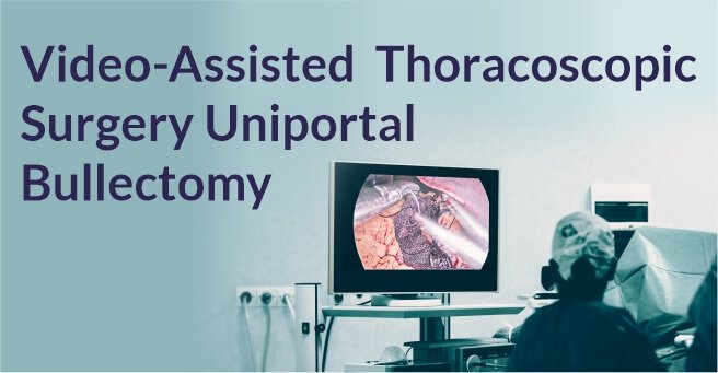
Video-Assisted Thoracoscopic Surgery Uniportal Bullectomy

Background

A 21 year old male patient presented with a history of dyspnea on exertion.

Diagnosis And Treatment

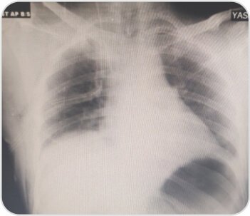
CXR showed right sided pneumothorax. ICD was placed. CT scan showed apical bullae. Uniportal bullectomy was done by Video-assisted thoracoscopic surgery (VATS).
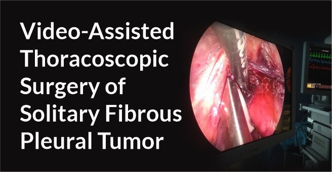
Video-Assisted Thoracoscopic Surgery of Solitary Fibrous Pleural TumorDr. BalasubramoniumMS (General Surgery), MCh(CVTS) Dr. Siva Prasad GoudMBBS, DNB (CVTS)Robotic & Minimally Invasive Thoracic Surgeon,Yashoda Hospitals, SecunderabadBACKGROUNDDIAGNOSIS & TREATMENTA 51 years old male patient came with a history of dyspnoea on exertion and purulent cough since 6 months. Pleural fluid aspiration was performed -6 times over 2 months. (1 - 1.5 litres of hemorrhagic pleural fluid was aspirated)Routine Investigations – WNLPFT:FVC – 1.26  FEV1 – 1.1  FEV1/FVC – 87.1ECHO – normal LV function , No RWMA  Pre procedure chest radiographs showingmassive right pleural effusionCT scan showing solitary fibrous right pleural tumor CTscanshowedtwolargeheterogenouslyenhancing pleural based soft tissue density lesions with neovascularity in right hemithorax measuring 14.3x13.4x11.5cm and  7.9x7.5x6.1cm.Surgery - Pleural aspiration was done and 2 litres of fluid was drained. Patient developed hemodynamic instability and was resuscitated in OT. Right posterolateral thoracotomy was performed and the tumor was removed under VATS.Post op chest X rayRight pleural tumorVideo-Assisted Thoracoscopic Surgery Uniportal Bullectomy

Video-Assisted Thoracoscopic Surgery of Solitary Fibrous Pleural TumorDr. BalasubramoniumMS (General Surgery), MCh(CVTS) Dr. Siva Prasad GoudMBBS, DNB (CVTS)Robotic & Minimally Invasive Thoracic Surgeon,Yashoda Hospitals, SecunderabadBACKGROUNDDIAGNOSIS & TREATMENTA 51 years old male patient came with a history of dyspnoea on exertion and purulent cough since 6 months. Pleural fluid aspiration was performed -6 times over 2 months. (1 - 1.5 litres of hemorrhagic pleural fluid was aspirated)Routine Investigations – WNLPFT:FVC – 1.26  FEV1 – 1.1  FEV1/FVC – 87.1ECHO – normal LV function , No RWMA  Pre procedure chest radiographs showingmassive right pleural effusionCT scan showing solitary fibrous right pleural tumor CTscanshowedtwolargeheterogenouslyenhancing pleural based soft tissue density lesions with neovascularity in right hemithorax measuring 14.3x13.4x11.5cm and  7.9x7.5x6.1cm.Surgery - Pleural aspiration was done and 2 litres of fluid was drained. Patient developed hemodynamic instability and was resuscitated in OT. Right posterolateral thoracotomy was performed and the tumor was removed under VATS.Post op chest X rayRight pleural tumorVideo-Assisted Thoracoscopic Surgery Uniportal Bullectomy

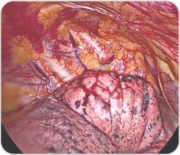
Right upper lobe apical segment bullae
Video-Assisted Thoracoscopic Surgery of Solitary Fibrous Pleural TumorDr. BalasubramoniumMS (General Surgery), MCh(CVTS) Dr. Siva Prasad GoudMBBS, DNB (CVTS)Robotic & Minimally Invasive Thoracic Surgeon,Yashoda Hospitals, SecunderabadBACKGROUNDDIAGNOSIS & TREATMENTA 51 years old male patient came with a history of dyspnoea on exertion and purulent cough since 6 months. Pleural fluid aspiration was performed -6 times over 2 months. (1 - 1.5 litres of hemorrhagic pleural fluid was aspirated)Routine Investigations – WNLPFT:FVC – 1.26  FEV1 – 1.1  FEV1/FVC – 87.1ECHO – normal LV function , No RWMA  Pre procedure chest radiographs showingmassive right pleural effusionCT scan showing solitary fibrous right pleural tumor CTscanshowedtwolargeheterogenouslyenhancing pleural based soft tissue density lesions with neovascularity in right hemithorax measuring 14.3x13.4x11.5cm and  7.9x7.5x6.1cm.Surgery - Pleural aspiration was done and 2 litres of fluid was drained. Patient developed hemodynamic instability and was resuscitated in OT. Right posterolateral thoracotomy was performed and the tumor was removed under VATS.Post op chest X rayRight pleural tumorVideo-Assisted Thoracoscopic Surgery Uniportal Bullectomy

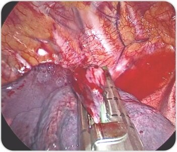
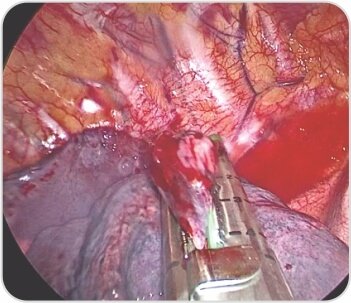
Bullectomy with endostaplers

Video-Assisted Thoracoscopic Surgery of Solitary Fibrous Pleural TumorDr. BalasubramoniumMS (General Surgery), MCh(CVTS) Dr. Siva Prasad GoudMBBS, DNB (CVTS)Robotic & Minimally Invasive Thoracic Surgeon,Yashoda Hospitals, SecunderabadBACKGROUNDDIAGNOSIS & TREATMENTA 51 years old male patient came with a history of dyspnoea on exertion and purulent cough since 6 months. Pleural fluid aspiration was performed -6 times over 2 months. (1 - 1.5 litres of hemorrhagic pleural fluid was aspirated)Routine Investigations – WNLPFT:FVC – 1.26  FEV1 – 1.1  FEV1/FVC – 87.1ECHO – normal LV function , No RWMA  Pre procedure chest radiographs showingmassive right pleural effusionCT scan showing solitary fibrous right pleural tumor CTscanshowedtwolargeheterogenouslyenhancing pleural based soft tissue density lesions with neovascularity in right hemithorax measuring 14.3x13.4x11.5cm and  7.9x7.5x6.1cm.Surgery - Pleural aspiration was done and 2 litres of fluid was drained. Patient developed hemodynamic instability and was resuscitated in OT. Right posterolateral thoracotomy was performed and the tumor was removed under VATS.Post op chest X rayRight pleural tumorVideo-Assisted Thoracoscopic Surgery Uniportal Bullectomy

Video-Assisted Thoracoscopic Surgery of Solitary Fibrous Pleural TumorDr. BalasubramoniumMS (General Surgery), MCh(CVTS) Dr. Siva Prasad GoudMBBS, DNB (CVTS)Robotic & Minimally Invasive Thoracic Surgeon,Yashoda Hospitals, SecunderabadBACKGROUNDDIAGNOSIS & TREATMENTA 51 years old male patient came with a history of dyspnoea on exertion and purulent cough since 6 months. Pleural fluid aspiration was performed -6 times over 2 months. (1 - 1.5 litres of hemorrhagic pleural fluid was aspirated)Routine Investigations – WNLPFT:FVC – 1.26  FEV1 – 1.1  FEV1/FVC – 87.1ECHO – normal LV function , No RWMA  Pre procedure chest radiographs showingmassive right pleural effusionCT scan showing solitary fibrous right pleural tumor CTscanshowedtwolargeheterogenouslyenhancing pleural based soft tissue density lesions with neovascularity in right hemithorax measuring 14.3x13.4x11.5cm and  7.9x7.5x6.1cm.Surgery - Pleural aspiration was done and 2 litres of fluid was drained. Patient developed hemodynamic instability and was resuscitated in OT. Right posterolateral thoracotomy was performed and the tumor was removed under VATS.Post op chest X rayRight pleural tumorVideo-Assisted Thoracoscopic Surgery Uniportal Bullectomy

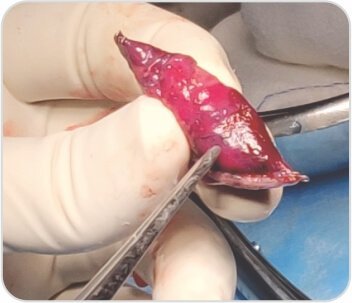
Excised bulla with portion of apical segment
Video-Assisted Thoracoscopic Surgery of Solitary Fibrous Pleural TumorDr. BalasubramoniumMS (General Surgery), MCh(CVTS) Dr. Siva Prasad GoudMBBS, DNB (CVTS)Robotic & Minimally Invasive Thoracic Surgeon,Yashoda Hospitals, SecunderabadBACKGROUNDDIAGNOSIS & TREATMENTA 51 years old male patient came with a history of dyspnoea on exertion and purulent cough since 6 months. Pleural fluid aspiration was performed -6 times over 2 months. (1 - 1.5 litres of hemorrhagic pleural fluid was aspirated)Routine Investigations – WNLPFT:FVC – 1.26  FEV1 – 1.1  FEV1/FVC – 87.1ECHO – normal LV function , No RWMA  Pre procedure chest radiographs showingmassive right pleural effusionCT scan showing solitary fibrous right pleural tumor CTscanshowedtwolargeheterogenouslyenhancing pleural based soft tissue density lesions with neovascularity in right hemithorax measuring 14.3x13.4x11.5cm and  7.9x7.5x6.1cm.Surgery - Pleural aspiration was done and 2 litres of fluid was drained. Patient developed hemodynamic instability and was resuscitated in OT. Right posterolateral thoracotomy was performed and the tumor was removed under VATS.Post op chest X rayRight pleural tumorVideo-Assisted Thoracoscopic Surgery Uniportal Bullectomy

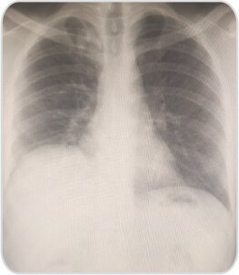
CXR at the time of discharge on POD 3

About Author –

Dr. Balasubramoniam K R, Consultant Minimally Invasive and Robotic Thoracic Surgeon, Yashoda Hospitals – Hyderabad
MS (General Surgery), MCh (CTVS)

About Author

Dr. Balasubramoniam K R | yashoda hospitals

Dr. Balasubramoniam K R

MS (General Surgery), MCh (CVTS)

Consultant Robotic and Minimally Invasive Thoracic Surgeon

About Author –

Dr. Siva Prasad Goud
MBBS, DNB (CVTS) Robotic & Minimally Invasive Thoracic Surgeon
Yashoda Hospitals, Secunderabad