
Robotic mediastinal tumor excision

Background
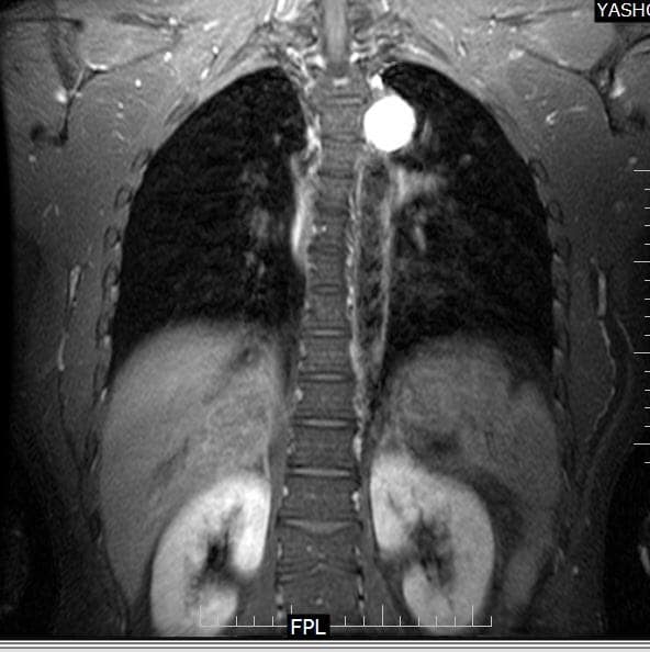
A 29-year-old male patient from Kerala was incidentally told about a left mediastinal tumor on a chest x-ray while undergoing screening for a job.
Diagnosis and Treatment
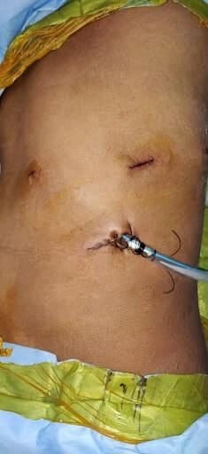
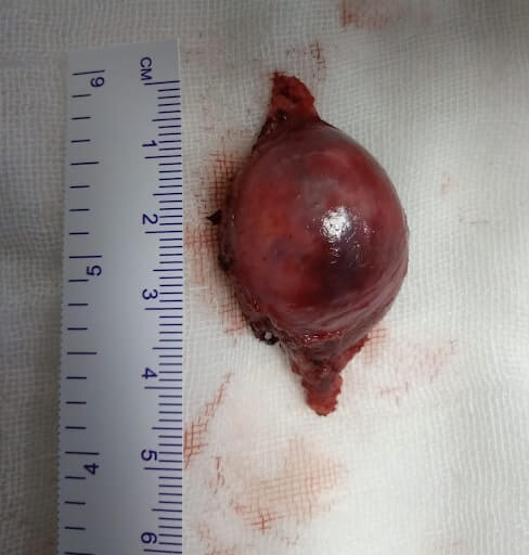
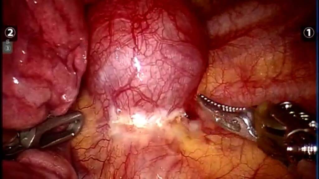
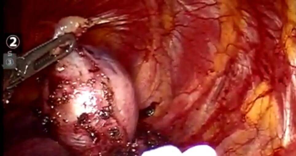
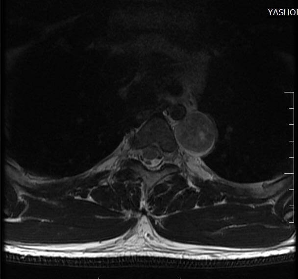
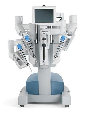
There were no comorbidities. The CT chest showed 3.7*2.7*2.6 cm smooth hypodense lesion in left paravertebral area at T3-T4 level. The MRI spine showed no spinal cord extension. The patient was taken for robotic mediastinal tumor excision. Histopathology report of the excised tumor suggested benign spindle cell lesion, probably of neural origin favouring schwannoma. The patient was discharged on the second postoperative day (POD 2).
About Author –
Dr. Balasubramoniam K R, Consultant Minimally Invasive and Robotic Thoracic Surgeon, Yashoda Hospitals – Hyderabad
MS (General Surgery), MCh (CTVS)
About Author

Dr. Balasubramoniam K R
MS (General Surgery), MCh (CVTS)Consultant Robotic and Minimally Invasive Thoracic Surgeon


 Appointment
 Second Opinion
 WhatsApp
 Call
Call More
More


