Giant Splenic Artery Aneurysm Treated by (Spleen Sparing) Endovascular Embolization

Background
A 48 year old female presented with dull aching epigastric pain which aggravated since last 4 days.
Diagnosis & Treatment
On evaluation, USG abdomen revealed an aneurysm in relation to splenic artery. CECT abdomen with CT angiogram revealed a large fusiform true aneurysm of size 5.5cmx 5cmx 4.5cm in relation to proximal and mid part of splenic artery.
The aneurysm was treated with Endovascular technique. The distal outflow of the aneurysm was embolized by coils followed by occlusion of proximal flow into the aneurysm by placing Amplatzer vascular plug device in the proximal splenic artery. Check DSA revealed complete occlusion of the aneurysm with preserved flow into distal splenic artery and splenic perfusion via gastroepiploic and short gastric arteries. On Post-operative day 1, Doppler study revealed completely thrombosed aneurysm. The patient was discharged in pain-free state without any need for splenectomy.
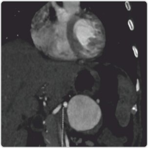
CT coronal reconstructed MIP image showing origin of aneurysm from splenic artery
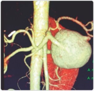
VRT image showing a giant fusiform splenic artery aneurysm
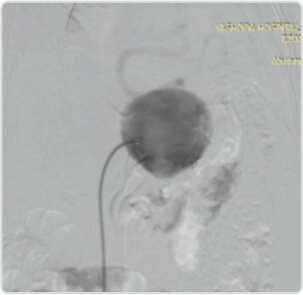
Pre embolization DSA image of splenic artery aneurysm
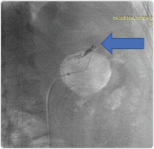
Fluoro image after distal coil (arrow) embolization of the outflow of aneurysm
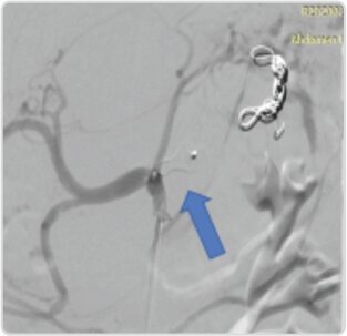
Complete occlusion of aneurysm after deployment of amplatzer vascular plug (arrow) in proximal splenic artery
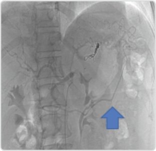
Post embolization distal splenic artery is being filled from Gastroepiploic artery (arrow)
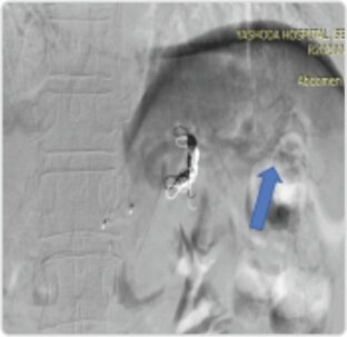
Post embolization of aneurysm, splenic perfusion is preserved
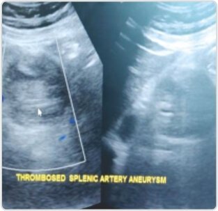
Follow up Doppler scan post-operative day 1 revealed completely thrombosed aneurysm
About Author –
Dr. Suresh Giragani, Consultant Neuro & Interventional Radiologist, Yashoda Hospitals – Hyderabad
MD (Radiology), DM (Neuroradiology)
Specialized in the comprehensive and widest range of vascular interventions covering neuro interventions, hepatobiliary interventions, venous, peripheral vascular interventions and interventions in cancer care.













 Appointment
 WhatsApp
 Call
Call More
More