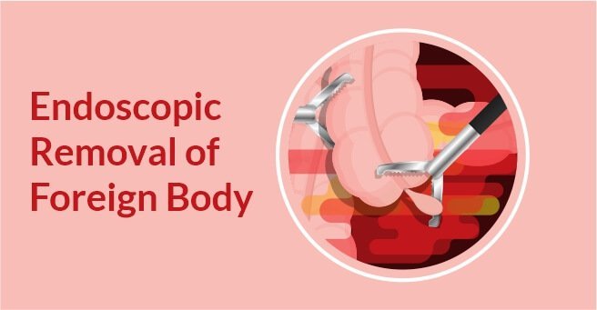
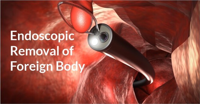
Endoscopic Removal of Foreign Body Case-2
Background A 3-year-old child presented to us 6 hours after ingestion of a sharp screw. Diagnosis and Treatment Abdominal X-Ray revealed the screw in duodenum. On endoscopy, the screw was identified in the second part of duodenum. It was repositioned into...
Continue reading...Endoscopic Removal of Foreign Body Case-1
Background A 54-year-old woman presented with dysphagia after meat ingestion. Diagnosis and Treatment On Contrast Enhanced Computed Tomography (CECT), bony fragment with sharp edges impacted in the esophageal wall was found just beyond the Upper Esophageal...
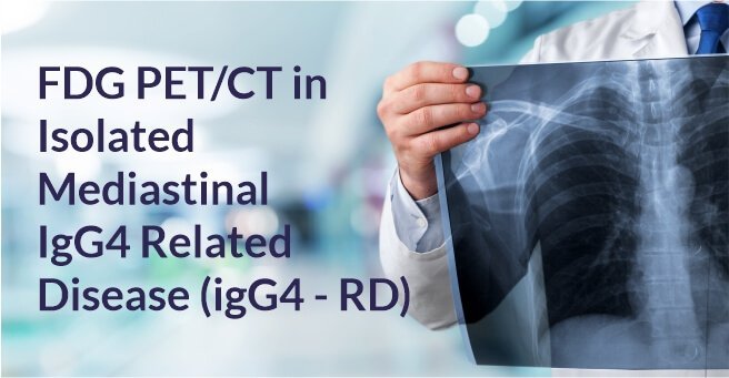
Continue reading...FDG PET/CT in Isolated Mediastinal IgG4 Related Disease
Background A 33 year old man on routine annual health check-up was found to have an opacity in the right suprahilar region on chest X-ray. Diagnosis CECT thorax done for further evaluation revealed soft tissue mass in the mediastinum. Mediastinoscopy and...
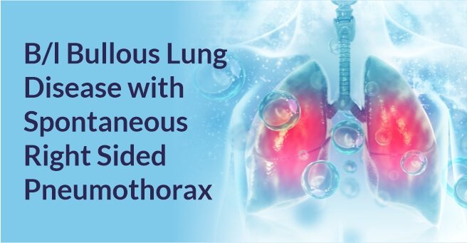
Continue reading...B/l Bullous Lung Disease with Spontaneous Right Sided Pneumothorax
Background 66 years old female patient presented with symptoms of shortness of breath, grade II to grade III since 2-3 months exacerbated since day 1 (grade IV). Patient is a known hypertensive and hypothyroid Diagnosis And Treatment On initial evaluation,...
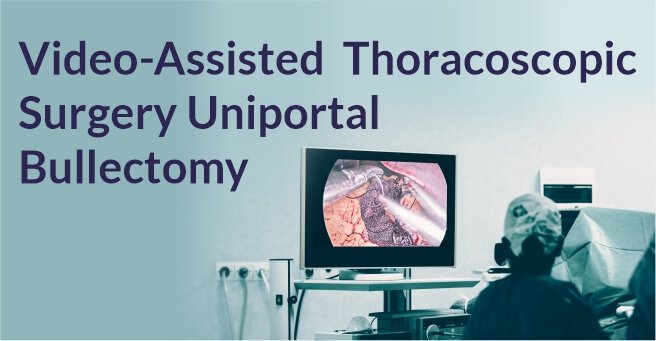
Continue reading...Video-Assisted Thoracoscopic Surgery Uniportal Bullectomy
Background A 21 year old male patient presented with a history of dyspnea on exertion. Diagnosis And Treatment CXR showed right sided pneumothorax. ICD was placed. CT scan showed apical bullae. Uniportal bullectomy was done by Video-assisted thoracoscopic...
Continue reading...



 Appointment
 WhatsApp
 Call
Call More
More

