Yashoda Hospitals > News > 31-year-old Woman Undergoes an Epidermoid Cysts Surgery at Yashoda Hospitals
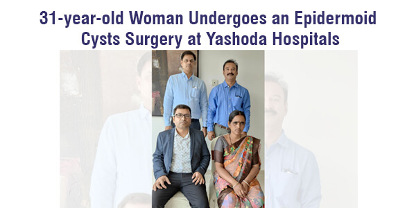
31-year-old Woman Undergoes an Epidermoid Cysts Surgery at Yashoda Hospitals


The MRI revealed the growth, which was extending forward up to the front of the midbrain and growing in the groove between the central brainstem and Cerebellum.
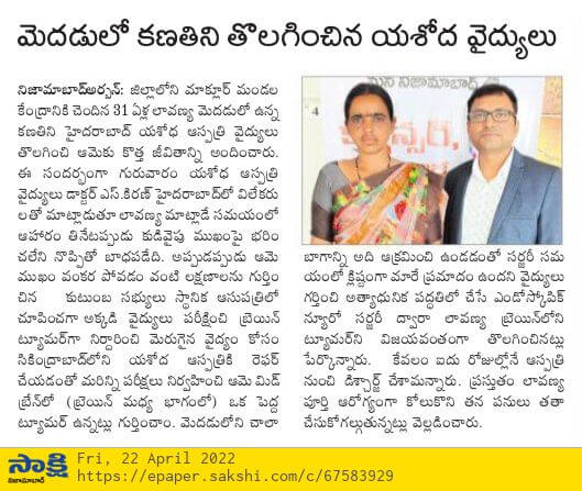
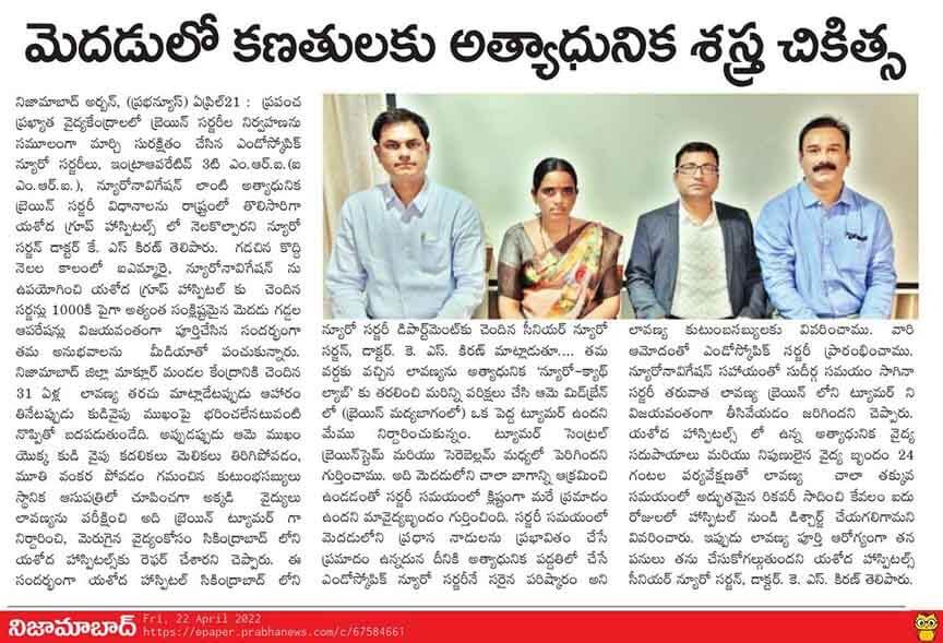
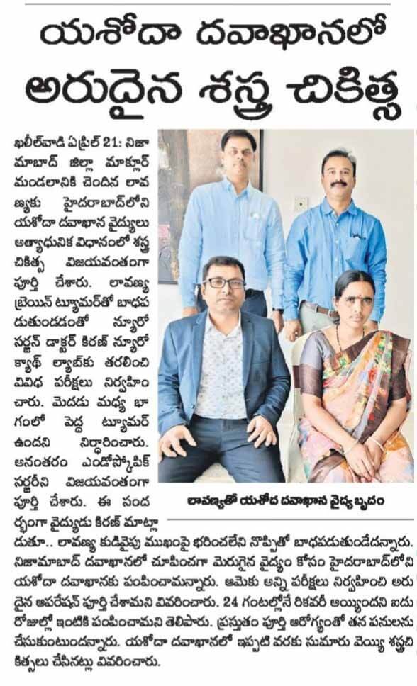
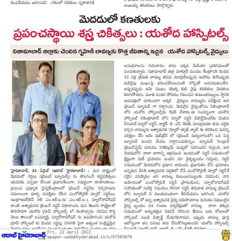
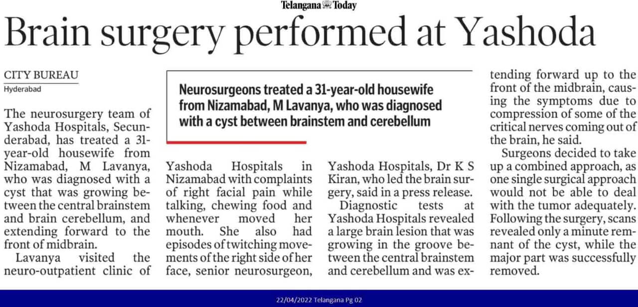
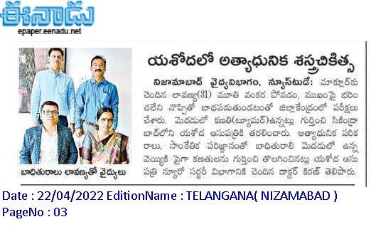
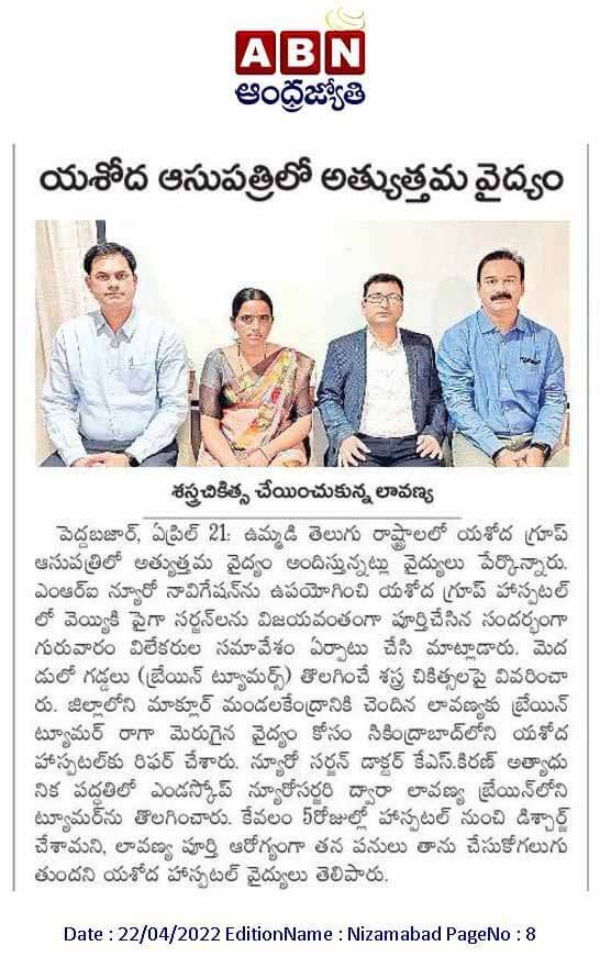
Nizamabad, 21st April, 2022: The neurosurgery team of Yashoda Hospitals Secunderabad has successfully performed a brain surgery on a 31 year old woman from Nizamabad, M.Lavanya who was diagnosed with epidermoid cysts in cerebellopontine angle.
Lavanya visited the neuro-outpatient clinic of Yashoda Clinic in Nizamabad with complaints of right facial pain that had been increasing when she talked, chewed her food, and made mouth movements for the past three months. She also had episodes of twitching movements of the right side of her face occasionally during this time.
Through clinical examination and MRI scan, Yashoda Hospitals neurologists discovered involvement of multiple nerves arising from the brain and deduced that there was a problem near the midbrain and the Cerebellum, which is located behind the brain. The MRI revealed the growth, which was extending forward up to the front of the midbrain and growing in the groove between the central brainstem and Cerebellum.
Dr K S Kiran, Consultant Neurosurgeon, Yashoda Hospitals, who led the brain surgery along with a team of neurologists, says, “This was a large brain lesion that extended from the right posterior region to the central brain region, causing symptoms due to compression of some of the brain’s critical nerves. We planned for a combined surgical approach after thoroughly analyzing the patient, as a single approach would not be able to deal with the tumor adequately.”
After counselling the patient about the procedure, which involves opening a small portion of the bone on the right side of the head behind the ear, a common approach to many such brain tumours, the brain surgery was performed. A high magnification microscope was used to remove as much of the tumour from the groove as possible, followed by the use of an endoscope to reach the part of the tumour deeper in the brain and remove a significant portion of the remnant tumour.
The patient was kept under observation after surgery and was given adequate fluids and antibiotics as needed. On follow-up, the patient had mild right-sided face weakness and an occasional cough, which gradually resolved.


 Appointment
 Second Opinion
 WhatsApp
 Call
Call More
More


